This is an online E log book to
discuss our patient's de-identified
health data shared after taking
his /her/Guardian's signed informed
consent.
Here, we discuss our individual
Patient's problems through series of
inputs from available global online
Community of experts with an aim to
solve those Patient's clinical
problems with collective current
evidence based inputs.
This E log also represents my
patient centered online learning
portfolio and valuable inputs on
Comment box is welcome.
Case Discussion :
I have been given this case to solve in an order to attempt to understand the topic of PATIENT CLINICAL DATA ANALYSIS to develop my competency in reading and comprehending clinical data including History, Clinical findings, Investigations and come up with a Diagnosis and Treatment plan.
A 32 year old female came to the OPD with the chief complaints of
* PAIN IN MULTIPLE JOINTS SINCE 1 YEAR
* FEVER AND COUGH SINCE YESTERDAY


HISTORY OF PRESENT ILLNESS:
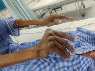
* Patient was apparently asymptomatic 1 year ago then she experienced pain in the proximal inter-phalangeal joints and later on to meta carpal joints and then to wrist, elbow, shoulder, knee, ankle too, on asking for order of involvement, she couldn’t recollect.
* Then she noticed flexion deformity of Metacarpo - phalangeal joints and swelling of the metacarpophalanheal joints over the course of illness
* She has to wait for 10 mins after waking up in the morning but the pain doesn’t subside and has to to continue to do the household chores.
* She went to nearby hospital after one month of initiation of pain and swellings and used medication for one month and on no improvement discontinued.
* She goes on and off to local doctors for pain of multiple joints almost of 8-10 times in the past 8 months which were giving her only temporary relief, on asking further she said that she has dryness of tongue on and off and burning sensation of eyes.
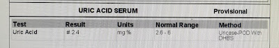
* She was admitted for 2 days in view of arthralgia and deformity of fingers with lab reports showing RA factor of 81 and ESR of 80.
* 4 days back around 7-8 pm in the night she noticed sudden weakness of right upper limb and left lower limb and was taken to near by hospital diagnosed to be hypokalemic with a potassium of 1.25 and was over corrected to 6.2 and after recovery, they were referred to Hyderabad I/v/o rheumatologist for joint pains, instead they came to KIMS and was advised on admission but as they weren’t prepared, they were not willing to admit.
* Since yesterday night she developed fever, insidious in onset, intermittent type, high grade with diurnal variation ( she has fever now too with a temp of 102.7F ) associated with dry cough.
HISTORY OF PAST ILLNESS:
* No History of Similar Complaints in the Past.
* Not a Known Case of DM, TB , Asthma, Epilepsy, Coronary Artery disease
PERSONAL HISTORY:
43 year old female who is married, Daily wage labourer
Diet - Mixed
Appetite - Normal
Sleep - Decreased
Bowel movements - Regular
Bladder Movements - Regular
Addictions - None
FAMILY HISTORY:
Not significant
GENERAL EXAMINATION:
* Patient is conscious, coherent and co-operative and lying on the bed.
* She is well oriented to time, place and person.
* She is mildly nourished, Thin built ( Cachexia )


Pallor - Absent

Icterus - Absent
Clubbing - Absent
Cyanosis - Absent
Lymphadenopathy - BILATERAL CERVICAL LYMPHADENOPATHY AND BILATERAL INGUINAL LYMPHADENOPATHY
Edema - Absent
VITALS AT THE TIME OF ADMISSION:
Temperature - 102 F
Pulse Rate - 89 BPM
Blood Pressure - 110/70 mm Hg
Respiratory Rate - 34 CPM
SPO2 - 98% at Room Air
Random Blood Sugar - 98 mg/dl
SYSTEMIC EXAMINATION :;
CARDIOVASCULAR SYSTEM - S1 and S2 present, No murmurs, Apex localised to usual place, No parasternal pulsations and Parasternal Heave.
RESPIRATORY SYSTEM - BAE +, NVBS
PER ABDOMEN - Soft and Non tender, No organomegaly, Bowel sounds heard.
CENTRAL NERVOUS SYSTEM - E4V5M6, HMF +


Provisional Diagnosis :
Symmetrical Polyarthritis secondary to ?? Rheumatic or ?? Autoimmune with Hypokalaemia secondary to Very Distal Renal Tubular Acidosis
Treatment :
1. INJ. NEOMOL 1GM / IV /SOS
2. TAB. PCM 650 MG / PO /SOS
3. TEMP charting 4th Hourly
4. TAB. METHOTREXATE 7.5 MG /OD/ ONCE A WEEK (EVERY FRIDAY)
5. TAB. FOLIC ACID 5 MG / ONCE A WEEK ( EVERY SATURDAY)
10/09/2022 :
S: Fever subsided and swelling of multiple Joints present
O:
Patient is conscious,coherent and cooperative
BP - 100/60 mm Hg
PR - 82 bpm
RR- 16 cpm
Temp- AFEBRILE
spo2 - 99% on RA
CVS-S1,S2 +,no added sounds heard
R/S-BAE+,clear
P/A-soft , non tender
CNS-NAD
A : Autoimmune arthritis with Hypokalaemia secondary to ? Renal tubular acidosis
P :
1. INJ. NEOMOL 1GM / IV /SOS
2. TAB. PCM 650 MG / PO /SOS
3. TEMP charting 4th Hourly
4. TAB. METHOTREXATE 7.5 MG /OD/ ONCE A WEEK (EVERY FRIDAY)
5. TAB. FOLIC ACID 5 MG / ONCE A WEEK ( EVERY SATURDAY)
11/09/2022 :


S: Fever subsided and swelling of multiple Joints present
O:
Patient is conscious,coherent and cooperative
BP - 90/50 mm Hg
PR - 72 bpm
RR- 16 cpm
Temp- AFEBRILE
spo2 - 99% on RA
CVS-S1,S2 +,no added sounds heard
R/S-BAE+,clear
P/A-soft , non tender
CNS-NAD
A : Autoimmune arthritis with Hypokalaemic paralysis secondary to ? Renal tubular acidosis
P :
1. TAB. NODOSIS 500 MG / PO / BD
2. TAB. PCM 650 MG / PO /SOS
3. TEMP charting 4th Hourly
4. TAB. METHOTREXATE 7.5 MG /OD/ ONCE A WEEK (EVERY FRIDAY)
5. TAB. FOLIC ACID 5 MG / ONCE A WEEK ( EVERY SATURDAY)
6. SYP. POTKLOR 5 ml + 1 glass of water / PO / BD
7. IV FLUIDS NS 1 UNIT @ 100 ML/ HR
DISCHARGE SUMMARY
Diagnosis - Hypokalemia secondary to Distal renal tubular acidosis with autoimmune arthritis(? Jaccouds arthropathy? Rheumatoid arthritis) TMJ Arthritis
COURSE IN THE HOSPITAL - Patient was admitted with the above complaints and investigations were done showing mild hypokalemia,hyperchloremia,abg showing metabolic acidosis and urine ph:6,USG s/o left renal caliculi,24hr urine potassium 15.6(high/pseudonormal for the degree of hypokalemia) suggesting renal wasting of potassum:features suggestive of distal RTA
X rays s/o autoimmune arthritis?Jaccouds arthropathy? Rheumatoid arthritis .Keeping in mind the association of autoimmune disease (RA)and distal renal tubular acidosis patient is started on Tab Methotrexate 7.5mg once weekly,Tab Folic acid 5mg once weekly,Tab Nodosis 500mg BD,syrup potchlor 5ml+1glass water BD.Patient is adviced to regularly be in follow up with her serum potassium and bicarbonate values .OMFS (TMJ arthritis) and orthopedic referral was taken and advice followed
ADVICE AT DISCHARGE -
1. Tab methotrexate 7.5 mg OD weekly once on Fridays
2. Tab folic acid 5mg once daily every Saturday
3. Tab Nodosis 500mg PO TID
4. Syrup potchlor 5ml in 1glass water BD
5. Tab Flexon MR PO OD *2days
6. Cold fomentation of preauricular area
7. Cementation of the maxillary FDP
Hand surgeon opinion on Tuesday /Friday's for deformed MCP joints(in follow up with dept of orthopaedics)
OMFS review after 1 week in view of TMJ arthritis(In view of Cementation of maxillary FDP)
GM review on 15/09/2022 with serum electrolytes
DISCUSSION AROUND THIS PATIENT :
Never received methotrexate or steroids?
4 days back there’s documentation of HCQ 100mg sir
Apart from it they said they don’t remember and usually they discarded
Involving both cervical and inguinal LN..?? Where's the problem...?
We can get X-ray hands bilateral ap view to document erosions and start her on steroids and single DMARD Mtx.
Oh.. seems to be severely effected joints
Can this be distal RTA sir ??
ABG showing bicarb of 8
Left renal calculus
RA??
Also urine ph 6
But abg is showing HAGMA
139-(104+8.7)=26
Na 139
K 3.2
Cl 104
The study of intermediates and end-products of metabolism in the context of immune cell functions is an emerging field that has been termed immunometabolism (Pearce et al., 2013). It is now clear that molecules such as succinate, lactate, acetyl-CoA, fumarate are more than intermediate by-products in metabolic pathways as they function as signaling molecules capable of linking metabolic reprograming with immune and inflammatory responses in immunity, inflammation and cancer (Figure 1; Haas et al., 2016). Whether metabolic perturbations are causal or the effect of the disease and how they can impact on the prognosis of RA is an area of significant current research.
Nuclear magnetic resonance (NMR) spectroscopy–based metabolomics on serum and urine samples from people with RA has identified a metabolic signature of patients with active established RA which differs from that of healthy controls (Young et al., 2013). Among the metabolites investigated 3-hydroxybutyrate and lactate were much higher in RA than in the control group. In addition choline, lactate and low-density lipoprotein (LDL) lipids strongly correlated with CRP a marker of disease activity (Young et al., 2013). This evidence suggests that NMR could be used as a tool to predict the development of atherosclerosis and other metabolic complications often associated with inflammatory disease.
https://www.frontiersin.org/articles/10.3389/fphys.2020.00347/full
Points in favour of distal RTA
Abg showing acidosis
Autoimmune condition
Hypokalemia
Urine ph >5.5
Renal caliculi
And most importantly a similar case report that I have found
Renal tubular acidosis (RTA) is a disorder of renal acidification characterized by inability to acidify urine to pH <5.5 despite the presence of severe systemic metabolic acidosis and hypokalemia. Hypokalemia leads to acute-onset paralysis and may be a presenting manifestation of RTA. Its association with various autoimmune disease has been reported previously in published reports, but has not been much emphasized. We, hereby, report a case of RTA that presented during the flare of rheumatoid arthritis (RA). A 42-year-old female, a known case of RA for 5 years, presented with persistent joint pain for 1 week and acute-onset quadriparesis for 3 days. Primary investigations revealed hypokalemia with metabolic acidosis. She was managed conservatively with potassium supplements and bicarbonate supplements along with steroids and disease-modifying anti-rheumatic drugs. Such a presentation of renal tubular acidosis in a patient during the flare of rheumatoid arthritis is distinctly rare and previously unreported in published studies.
Phase 1 and 2 logic but no phase 3 experiments?
What is our current patient getting? Or are we waiting for her 24 hour electrolytes? Could you speak to Biochemistry madam?What is our current patient getting?
Only potassium and calcium?
Please get sodium and creatinine too
Not yet started on potassium or bicarb sir as she started collecting 24hr urine from today mrng.
I asked them sir ..they said they will do 24hr urine calcium also
I am not able to find phase 3 sir.
Can anyone help me with finding RCT/epidemiological study placebo vs bicarb in dRTA
Yes sir
24hr urine sodium,potassium,calcium, creatinine and protein
(As CUE showed alb+)
One reason is that phase 3 can be more challenging to do in rare disorders due to less sample size.
Yes sir
No consent given for amputation or debridement of the right lower limb.
https://www.ncbi.nlm.nih.gov/pmc/articles/PMC6152546/
Please see is there any lactic acidosis...??
I'm sorry, is it only me or does anyone else notice too that there are no erosions whatsoever? These are just joint subluxations?
It's there in the image shared by Chandana
A more lateral view is showing subluxation 😅
I could appreciate erosions too
Correct me if am wrong
This one? From Proximal to distal
Radius and Ulna look normal. Metal bangle visible on left forearm.
Carpal bones morphology and architecture preserved.
Radial deviation of all metacarpal bones with no erosions.
Ulnar deviation of all PIPs with no erosions I can see. Can someone please point out the erosions to me please?
Check the carpal bones again
Maybe a few erosions in the right hand 4th and 5th digits. RA is often worse in the dominant hand.
But nothing justifying her hand morphology.
How are the movements at the wrist?
Why is a joint exam not performed yet?
Done sir
No restriction of movement at joints
Apart from fixed flexion deformity at MCP
Bingo!! See how valuable the clinical exam is. So no Carpal bones erosion clinically then?
65 degrees and above is normal 👍🏽

Definition of bone erosion :
Bone erosion is a radiological term and reflects the fact that imaging is used for detection.10 Erosions are visible on plain radiographs as breaks in the cortical bone surface, and are often accompanied by loss of the adjacent trabecular bone."
https://www.ncbi.nlm.nih.gov/pmc/articles/PMC4096779/
Also show the flexion
Well done to all. Ideally a good start for rheum exam is the pGALS system.
Preliminary Gait, Arms, Legs and Spin
Of course yes sir. But I still can't appreciate them.
She is sleeping sir
Managed to get one arm
Again 65 and above is normal
https://www.ncbi.nlm.nih.gov/pmc/articles/PMC8497874/#!po=3.57143
Alkali therapy may be used to correct acidosis in patients with distal or proximal RTA .In patients with distal RTA, alkali therapy also corrects for hypokalemia. Alkali therapy with 1–2 mmol/kg/day NaHCO3 or KHCO3 is normally sufficient to equal daily acid production . however, in patients with nephrolithiasis or nephrocalcinosis, the elevated Na+ load with NaHCO3 therapy may cause increased urine calcium excretion, which can precipitate kidney stone formation. In these patients, K-citrate administration is preferable; this will also increase urine citrate excretion and prevent recurrence of kidney stones . Patients with severe hypokalemia should also receive K+ replacement (i.e., with KCl or K-citrate) to prevent further lowering of serum K+ concentrations and symptomatic hypokalemia . Long-term treatment of distal RTA generally requires a combination of NaHCO3 and KHCO3 . Children with distal RTA require sufficient NaHCO3 or KHCO3 (usually 4–8 mmol/kg/day) to maintain normal serum HCO3– concentrations and prevent growth retardation
https://link.springer.com/article/10.1007/s12325-020-01587-5
According to the patient, she can't open her mouth nore than this sir... If she opens , she is feeling pain in the left lumbar region
This was the reason
I put a pic of lower side of face
Skin is adherent to bones ??
On asking her husband he said it’s the same way since marriage even she denied any change in face
Asked for pics of childhood hopefully we get it tomorrow
But there was decreased laxity of skin proximal or distal parts
No decreased laxity of skin **
She has dental implantation ( upper four incisors ) surgery 10 years ago due to the cosmetic problem. No postsurgical infection and complications
Check out her left Tempomandibular joint in comparison to her right.
See if you can insert a gloved finger into her cheeks and compare the two sides for any induration differences. Get our current facio maxillary touring resident to do it also.
Found this past case which fortunately was done by our elective Intern @Sanjana Palakodeti here https://bmjsanjana.blogspot.com/2019/03/31-yr-old-female-come-to-hospital-with.html?m=1 on March 2019 when she visited and when Lakshmana 2019 PG took offline ownership.
After going through it I can guess where yesterday's collective cognition around the erosions had originated from @Aditya Samitinjay ?
***Discussion and questions in today mrng rounds
1. RA without carpel bone involvement
2. ?Jaccouds arthropathy ?RA ?SLE
https://jharipriya.blogspot.com/2022/09/this-is-online-elog-book-to-discuss-our.html
9.Deflazacort vs prednisolone in autoimmune arthritis
This description perfectly fits the differential your team is suggesting sir? @Rakesh Sir GM
https://www.ncbi.nlm.nih.gov/pmc/articles/PMC8497874/#!po=3.57143
So She might have had a history of RHD with no fever or a system involvement ??
Clinically I couldn’t find any murmurs or may be didn’t appreciate it
Was Echo done ?? @Dr. Haripriya Ma'am Med PG
Anion gap
143-(12.4+103)=27
Delta anion gap=(pt anion gap-normal anion gap)=27-10=17
Delta bicarb =difference between normal and pt bicarb=25-12.4=12.6
If delta anion gap Is more than delta bicarb It means the patient is having both normal and high anion gap
If delta anion gap = delta bicarb patient is only having high anion gap
What about potassium and bicarb in her treatment? 24 hour urinary results?
We dint start her yet on those medication sir.She is collecting her 24hr urine.will start them after she stops collection tomorrow at 8am
The pathophysiological mechanism of dRTA in relation to autoimmunity remains unclear. Several reports suggest that autoantibodies against the CAII enzyme [36, 57] or the acid–base transporters are involved in the pathogenesis of dRTA in autoimmune disease
https://www.ncbi.nlm.nih.gov/pmc/articles/PMC4107275/
24hr urine protein is 195 sir,urinary creatinine 0.6,1300ml volume.Can we still consider SLE and send testing for immunological criteria sir??fever (constitutional domaina) and joint involvement +.atleast one clinical criteria is required and total of 10 points is needed
Jaccouds is seen in SLE
Chasing that diagnosis won't change our management plan currently
https://www.nejm.org/doi/full/10.1056/NEJM196207052670103
Urine potassium15.6sir(25-125 mmol/lit is normal)🙆🏼♀️it came lower than normal sir😅
Urine sodium is 228 sir
Check if it can come low in distal RTA
Urine anion gap in our patient is positive(228+15.6-221=26) indicating RTA in our patient
Gastrointestinal bicarbonate losses can be differentiated from RTA by estimating the urine anion gap. Negative urine anion gap indicates increased renal NH4 + production (extrarenal cause for metabolic acidosis), while positive gap suggests RTA
https://www.google.com/url?sa=t&source=web&rct=j&url=https://med.stanford.edu/content/dam/sm/pednephrology/documents/secure/1Renaltubdisorders.pdf&ved=2ahUKEwjs7cTxuoz6AhX6H7cAHU0nCTUQFnoECAcQBg&usg=AOvVaw0DGihPwaEYFM4SkGxaki6r
I am not finding urine potassium seperately being tested in dRTA sir.its urine anion gap they are referring to most of the times
Can you check what is normal Urine K+ range? Standardized values and not this lab value?
For adults, normal urine potassium values are generally 20 mEq/L in a random urine sample and 25 to 125 mEq per day in a 24 hour collection.
https://www.ucsfhealth.org/medical-tests/potassium-urine-test#:~:text=Normal%20Results,of%20potassium%20in%20your%20body
This is because the urine K+ should have been much lower for that hypokalemia and this is classically "pseudonormal". Same like with normal PTH in hypercalcemia is abnormal

9/10, 8:11 PM] Rakesh Sir GM: What about her mouth opening? Did we get the OMFS opinion?


As perhaps mentioned above by earlier this is another consequence of her rheumatoid arthritis.
Hope our current case reporters clarify the association between RA with Jaccoud's arthropathy and distal RTA clearly in this case


No comments:
Post a Comment